|
|
|
The Collie is a relatively healthy breed and usually lives to an
average of 12 years old. We give a written health guarantee on all
of our dogs. Below you will find information on the care of the
Collie and diseases it can be prone to as well as links to other
sites of information.
On this page you will find information on Collie Eye Anomaly, Hip
Dysplaysia, Degenerative Myelopathy, MDR1 - Drug Sensitivity and The
Collie Health Foundation.
Collie Eye Anomaly
|
Eye Disease in Collies
"Your Collie's Eyes"

(Cross-section of the Canine Eye)
Like people, Dogs are subject to a large number of inherited
eye diseases, two of which can affect a Collie's eyes, CEA
and PRA, should be of concern to all breeders.
CEA
CEA is an inherited eye disease common to the Collie breeds
including the Rough Collie. In most cases the disease in
inherited in a very mild form, so mild in fact that
it cannot be detected by clinical examination, and in this
mild form it is not believed to affect vision at all. CEA is
not progressive, generally speaking what we see in an 8 week
old puppy will not worsen with age, except in rare cases
where large coloboma are present that can later cause
retinal detachment. In 75% of cases where CEA is diagnosed,
the degree of the disease is ‘mild’ and the dog will lead a
normal life. A percentage of those diagnosed as ‘affected’
will have one or more small coloboma, tiny holes or dents in
or near the optic disc. Provided the coloboma is small,
sight will not be affected to any huge degree and once again
the dog will lead a normal life.
We need to remember that dogs evolved to hunt prey at dawn
and are not nearly as dependent on vision as are humans.
Both scent and hearing are equally involved and it can be
said that a dog is only 33% dependent on sight.
Generally speaking therefore a small coloboma will not
greatly detract from the dog’s overall quality of life.
However in a very small percentage of cases that are
diagnosed with CEA, a large coloboma and or haemorrhage is
present, and in these instances it can lead to detached
retina. If the dog has a detached retina this will cause
blindness in the affected eye. Occasionally a puppy is born
with severe impairment of vision in both eyes and it is
possible to produce an entire litter thus affected, for this
reason all puppies should be eye tested between the age of
6-8 weeks.
http://www.laboklin.de/index.php?link=labogen/pages/html/en/geneticdiseases/dog/dog_cea_collie.htm
Many puppies diagnosed age 6-12 weeks as ‘mildly affected’
will appear to be completely free of CEA if tested when
older. This phenomenon is known as CEA ‘go-normal’ although
it must be remembered that from a genetic viewpoint, such
dogs ARE affected.
For many years breeders have tried to eliminate CEA or at
least reduce the incidence and severity. However all efforts
have failed and the occurrence and severity of CEA are much
the same now as they were when the condition was first
diagnosed in the breed. This has confused those breeders
with an understanding of basic genetics. CEA is inherited as
an autosomal recessive trait. Both parents must carry at
least one copy of the defective gene for the offspring to be
affected. If those collies diagnosed CEA clear under
clinical examination at 6-8 weeks are actually ‘clear’ the
following should be applicable.
First of all I shall be referring to CEA non carriers as
+/+, CEA clear/carrier as +/-, and affected as -/-
We need to remember that ‘mildly affected’ shares exactly
the same gene responsible for producing ‘severely affected’
puppies although it is believed a dog inherits certain
'modifier' genes seperately which do affect the severity of
the diseases. Even so, CEA is one gene that displays itself
with varying severity in individual puppies and coloboma are
part and parcel of the SAME gene.
+/+ mated with +/+ will produce 100% +/+ puppies.
+/+ mated with +/- will produce 50% +/+ puppies and 50% +/-
puppies
+/+ mated with -/- will produce 100% +/- puppies.
+/- mated with +/- will produce 25% +/+ puppies, 25% -/-
puppies, 50% +/- puppies.
-/- mated with -/- will produce 100% -/- puppies.
The question for those breeders genuinely interested has
been...why have no CEA non carriers been found amongst the
UK Collie population?, if the puppies clinically diagnosed
as clear are truly either +/+ or +/- it would be genetically
impossible NOT to produce a percentage of non
carriers. So we must ask ourselves...where are these non
carrier Collies? Surely they can’t ALL be hidden from the
breeding gene pool and in pet homes!
In recent years a new form of diagnosis has been developed,
the canine genome has been mapped and a DNA marker found for
CEA. At long last an answer to our question is being
revealed. Several UK bred Collies living both here in the UK
and also on the European continent have now been DNA tested.
To my present knowledge every UK bred Collie tested by DNA
analysis has been diagnosed as ‘affected’ and not a single
collie has been diagnosed +/- or +/+. The Collies used in
these tests had all been diagnosed at 6-8 weeks by clinical
analysis as CLEAR....but in reality they are AFFECTED. This
fully explains why breeders have been unable to make drastic
improvements in this area in the UK, and it is extremely
possible we have NO ‘true’ clear eye gene in the UK Rough
Collie population. Clinical examination has proven to be
untrustworthy, although severity of the condition can be
discovered by this method.
Indeed, world wide it is believed that as few as 5% of Rough
Collies are CEA non carrier (+/+) with perhaps 15% worldwide
being clear/carrier (+/-) and as I have already said, No
true clears have yet to be discovered in the UK...that is
until now! We recently imported two CEA non carrier (+/+)
dogs from Canada and are integrating them into the gene pool
here at Wicani, every puppy sired by these boys is +/- (by
DNA) making collie history here in the UK, as these are the
only UK bred puppies presently known to carry the TRUE clear
eye gene! A third non carrier Collie has been imported to
the Strobroy kennel in Scotland.
We plan to import several more Collies to further enhance
our gene pool, increasing genetic diversity whilst
introducing a true DNA marked CEA free gene. I feel humbled
and honoured to be in a position whereby I am able help the
breed in this way.
I am in no way criticising my fellow breeders that have made
different decisions and prioritised differently in their
breeding programs, and as I have already said, a collie
affected with CEA seldom shows signs of impaired vision and
many will appear to be clear of this condition in clinical
examination. Another point worth remembering is that the
Breed has lived with CEA since its beginnings, however, on a
personal note I wish to begin the eradication of any
defective gene wherever possible, and if I can prevent the
birth of one blind puppy, I feel my task will have proven
itself worthwhile.
For those interested I use in my home pages the following
terminology:
NORMAL EYED NON CARRIER (+/+) A Collie that is not carrying
the gene for CEA and cannot pass an affected gene to their
offspring
(also referred to as N/E N/C)
NORMAL EYED (+/-) A Collie with the true clear eye gene but
carrying an ‘affected’ gene. This Collie can pass to its
offspring either a true clear gene or an affected gene.
(also referred to as N/E)
CLINICALLY CLEAR (-/-) A Collie that has been diagnosed
‘clear’ by clinical examination, but if DNA tested would
prove to be ‘affected’ this Collie can only pass an affected
gene to its offspring, even though he/she ‘appears’ to be
clear themselves and even if the offspring ‘appear’ to be
clear by clinical examination.(also referred to as C/free)
AFFECTED (-/-) A Collie who appears ‘affected’ (whether mild
or severe) on clinical examination.
REMEMBER, the only TRUE way to check a Collies eye status is
by DNA analysis.
Contribuition:
Angela Harvey. (Wicani Collies)
PROGRESSIVE RETINAL ATROPHY
The other Collie eye problem that can occur in rare
instances is Progressive Retinal Atrophy (PRA). Since
the name is just what it implies, it can be a progressive
disease, that may not appear until later in life. This is a
completely different and unrelated disease to CEA. As the
name indicates, PRA is a progressive disease which refers to
retinal degeneration. It can result in complete blindness in
one or both eyes. However, Collies seem to be blessed with
the fact that PRA seems to have an early onset. Fortunately,
this is an eye disease that has largely been eradicated
thanks to breeders efforts of test breeding potential
carriers. Since PRA is a simple recessive gene, it is much
easier to test for than CEA. Also, thanks to funding of
certain grants by the Collie Health Foundation, research is
being done to locate the genetic markers for this disease,
which will further reduce the occurrence. Currently there is
already a genetic test for PRA, please consult OPTIGEN
website for more details!
PRA has proven to be a simple recessive in all the breeds
studied. Again, this means that even though the condition is
not present at birth, both parents must be
carriers. If one parent has PRA, half the puppies may
develop PRA, but all will be carriers for the disease. Early
signs of the problem may be noticed by the owner as "night
blindness." The dog has trouble seeing in dim light and will
bump things. An expert may detect early signs in the eye at
six months or younger.
For more info:
..\Collie Eye
Anomaly.doc |
Drug Sensitivity - MDR1 GENE
MDR1(-/-) THE SILENT KILLER
Liver failure, fading puppies, small litters, could this gene
mutation be responsible....????
By Angela Harvey - Wicani Collies
Most people involved in our Breed know and appreciate that some
Collies are sensitive to certain Drugs, the common thoughts with
regard to
this being that provided we stay clear
of all known at risk drug, our Collies will carry on living long
healthy normal lives simple.?
The problem was first discovered quite by accident when researchers
experimented on laboratory mice, the mdr1 protein is one of the
things separating mammals from insects and bugs, and as such is
present in all mammals including man. Researchers were interested to
learn what would happen (if anything) if this protein were absent.
To this end an experiment was set up and the mdr1 protein was
removed from a family of mice. For many months these mice lived an
entirely normal existence, eating, sleeping, mating and rearing
their young, researchers began to think the missing MDR1protein was
making no difference at all to the lives of these mice, until the
mice developed a mite infestation. The cages were sprayed with
Ivermectin, the following day every mouse was dead.
Since this time we have discovered many drugs fatal to collies
carrying the double MDR1 gene mutation (-/-).
For years I have kept a private data base of collies having died of
liver and kidney problems, having experienced liver problems myself
in the past, I wanted to know if other dogs dying with these
problems were related to those I had lost myself. Over the years the
data base grew and remained quite confusing, that is until we
discovered the MDR1 problem. As people began to make public the MDR1
status of their dogs I began to notice a pattern emerging. The lines
commonly found to contain a large number of (-/-) dogs, were in fact
often the same lines from which the dogs in my data base were bred.
More recently, some dogs have died after having been tested for the
MDR1 mutation, to press these have all been -/- , food for thought!
Plus, when it was discovered a line free of liver problems, and
included it into Wicani´s breeding programme, not only did she got
her dogs rid of liver problems, but when they were tested for MDR1,
they were +/+ in other words they were free of the mutation. This
could be coincidence, so Angela began research into what happens
when the MDR1 protein is absent in humans.
It seemed common sense to her, that if poisons and chemicals were
crossing the blood brain barrier and entering the brain, surely
lesser toxins were doing this all the time but not to an immediately
fatal degree. As the MDR1 protein is responsible for pumping these
toxins away from the brain and out of the system, could these toxins
be remaining in the body and being stored in the liver? What about
the toxins and chemicals normally passed through the dogs body from
complete diets, travel sickness pills etc; were these being stored
in body organs, building up over time to create problems? If I was
correct with her theory, the result would be fabulous, It would mean
the final solution is within our grasp to rid our breed of some of
the persistent liver and kidney related problems that have plagued
us.
Angela´s research into humans revealed some interesting facts, one
being that when the MDR1 P-glycoprotein is absent, the placenta
works differently. Poisons, lesser toxins and even some viruses not
only cross the blood brain barrier; they also cross the placenta
when they would not normally do so. Such humans often suffer with
Colitis too.ring any bells yet?
For a long time she wondered how research into this gene mutation in
Collies could be funded, my prayers have been answered.
Giessen
University have now done
several studies, the results are proving to be very interesting.
Steroids like Cortisol are also transported by the P-glycoprotein
(this is the protein that cannot be produced by MDR1 -/- dogs) a new
study has now been done in this area. One thing quickly became
apparent, In MDR1 -/- dogs there is a lower level of Cortisol in the
body, predisposing such dogs to greater problems when under stress.
It would appear that MDR1 dogs really do suffer more stress and
stress related illnesses. Other revelations presented by Professor
Dr. Geyer of the University are, the placenta works differently when
the bitch is MDR1 -/-and yes, toxins, viruses and chemicals do cross
the placental barrier in bitches and not only humans. There are now
at least 100 substances known to be dangerous to the MDR1 double
mutant dog, and the list is growing. The fact that such dogs have a
huge improvement in health when fed a natural raw meat diet
emphasises the possible problems with toxin overload when fed a
modern complete diet. In MDR1 -/- dogs, antibiotics are far more
dangerous. Most people never consider antibiotics to be poisonous
but they ARE they are designed to poison bacteria.
Certain Antibiotics can destroy the liver of a double mutant dog
within days..!!!! If your dog is in this category, and needs such
medication, ask your vet to do blood tests at regular intervals
throughout the treatment to ensure no irreversible damage is being
done. Antibiotics or Steroids should NOT continue more than one
week, and if they must, blood tests must also be done. Many Breeders
presently have a policy of giving antibiotics randomly to bitches
when mated, in light of this latest research is this really wise?
Could this be one of the reasons some bitches are dying of liver
failure shortly after whelping and could it responsible for ever
decreasing litter sizes? Unless you know the status of your bitch,
you could be poisoning her and possibly her puppies too!
MDR1 protein begins working when food or medicines enter the
stomach. Many things are transported out when the dog is MDR1 +/+,
but when the dog is MDR1 -/- the entire dosage enters the
blood stream, where it is transported not only directly to the
brain, but to every other organ of the body. They enter organ cells
and the placenta of developing embryo where they remain for far too
long.
Another big problem revealed itself. If an MDR1 -/- dog is given a
cocktail of anaesthesia AND antibiotics together, it can totally
destroy the liver! When a bitch is spayed, such procedure is
normal, how many collie bitches have died or been diagnosed with
liver failure within a short time of being spayed?
In the past we knew nothing about the MDR1 P-glycoprotein, but now
we do. In her opinion it is the single most important problem within
our Breed, but the good news is WE CAN BREED IT OUT. Unlike CEA (in
the UK
we presently have no known genetically clear eyed collies) and Hip
Dysplasia (which I believe is polygenetic and influenced by
environmental factors as well as genetic), MDR1 can be
eradicated easily, and if we love the Breed, we owe it this much.
Can we really continue breeding animals knowing they are or could be
failing in this respect?
Perhaps we could begin by testing our dogs, and making those results
known to all fellow breeders. Perhaps if our stud dog is -/- we
should refuse bitches to him unless they are +/+ Likewise if your
bitch is -/-, wouldnt it be wise to find her a partner who is +/+?
Perhaps the next time you have a litter of puppies born and are
debating which to keep because you particularly like two bitches�have
them MDR1 checked and let the result decide. Slowly we can move
forward.
Angela owned her first show collie in 1974; She began studying the
Breed in 1972. Rough collies have brought so much joy into her life.
We live in exciting times; we live in a time when we can give
something back to the Breed. In her opinion the missing MDR1
P-glycoprotein is the silent killer, being aware of every dogs
status is one step closer to life.
The following links are recommended.
http://www.pubmedcentral.nih.gov/articlerender.fcgi?artid=1636591
http://www.pubmedcentral.nih.gov/articlerender.fcgi?artid=1636591&rendertype=table&id=t1-cvj47pg1165
http://www.vetmed.wsu.edu/depts-VCPL/genetics.aspx
http://www.vetmed.uni-giessen.de/pharmtox/juniorprof/research.php#res_02
http://www.scielo.br/scielo.php?pid=S0103-84782006000100056&script=sci_arttext
Contribuition:
Angela Harvey. (Wicani Collies)
______________________________________________________________________________________________________________________
Hip Dysplaysia
|
OFA stands for Orthopedic Foundation for Animals. It is a
"not for profit organization", with the purpose to "provide
a standardized evaluation for hip dysplasia and to serve as
a data base for control of hip dysplasia through selective
breeding." In order to receive an OFA number, a dog has to
be at least 24 months or older on the day of his X-rays.
Younger dogs can be x-rayed and evaluated but cannot receive
an OFA number. Many breeders do this as a potential early
screening. Because of the difficult positioning of the rear
legs, (they must be extended and pulled parallel), most dogs
require sedation or anesthetic. Film identification is
extremely important. Permanent film identification in the
film emulsion is required for all radiographs. Upon
completion of X-rays, the owner fills out an OFA
application. The radiograph, signed application and fee are
then submitted to OFA. OFA also recommends that a copy of a
dog's AKC registration be enclosed.
Once the x-rays are received by OFA the process first begins
by screening the X-rays for correct positioning and
technique. If acceptable, the X-rays are then evaluated by
(3) board certified Veterinary radiologists and a consensus
of their opinions is taken. "The hips are evaluated for
subluxation, shallow acetabulum (socket), femoral head/neck
remodeling, acetabular rim/edge changes and degenerative
joint diseases."
THE GRADING CATEGORIES ARE:
Excellent, good and fair....all
considered Normal and will receive OFA numbers.
Borderline......Recommend a recheck in 6-8 months.
Mild, Moderate and severe...Dysplastic.
As with CERF, in order for the OFA number to be on the dog's
AKC registration form, as of July 1, 1996, a dog must be
either tattooed or micro-chipped at the time the X-rays are
taken. This identification should be noted on the X-rays.
OFA sends a quarterly report of OFA numbers to AKC.
The OFA number is similar to the CERF number, in that each
element has a precise meaning. Using the number...
CO-1620E24M-T as an example..CO is the breed identifier (in
this case for Collie); 1620 is the ascending numerical order
of normal individuals assigned a breed registry number; E
stands for Excellent; 24 is the age in months when the
x-rays were taken; M is for the sex of the dog and T stands
for tattoo. An OFA number is good for the entire lifetime of
the dog, but OFA reserves the right to correct or revoke any
number.
Of added interest, OFA is branching out into other areas of
health, with the recent addition of a Canine Thyroid
Registry and a registry for Congenital Heart Disease as of
January1, 1996.
Further information on OFA may be obtained by writing or
calling: OFA at 2300 E. Nifong Blvd, Columbia MO 65201-0418,
telephone 1-573-442-0418; Also information may be obtained
from The American Kennel Club, at 1-919-233-9767. Here is a
link to the
OFA website:
Written by Gayle Kaye - from the April 1998 CCA Bulletin.
|
Degenerative Myelopathy - Disease Basics
|
http://www.cvm.missouri.edu/neurology/dm/index.html |
|
What is Degenerative Myelopathy?
Degenerative myelopathy is a progressive disease of the
spinal cord in older dogs. The disease has an insidious
onset typically between 8 and 14 years of age. It begins
with a loss of coordination (ataxia) in the hind limbs.
The affected dog will wobble when walking, knuckle over
or drag the feet. This can first occur in one hind limb
and then affect the other. As the disease progresses,
the limbs become weak and the dog begins to buckle and
has difficulty standing. The weakness gets progressively
worse until the dog is unable to walk. The clinical
course can range from 6 months to 1 year before dogs
become paraplegic. If signs progress for a longer period
of time, loss of urinary and fecal continence may occur
and eventually weakness will develop in the front limbs.
Another key feature of DM is that it is not a painful
disease.
|
Degenerative myelopathy is a devastating disease causing
progressive paralysis in a large number of dog breeds.
New research has identified a gene that is associated
with a major increase in risk of the disease.
|
|
What causes Degenerative Myelopathy?
Degenerative myelopathy begins with the spinal cord in
the thoracic (chest) region. If we look under the
microscope at that area of the cord from a dog that has
died from DM, we see degeneration of the white matter of
the spinal cord. The white matter contains fibers that
transmit movement commands from the brain to the limbs
and sensory information from the limbs to the brain.
|
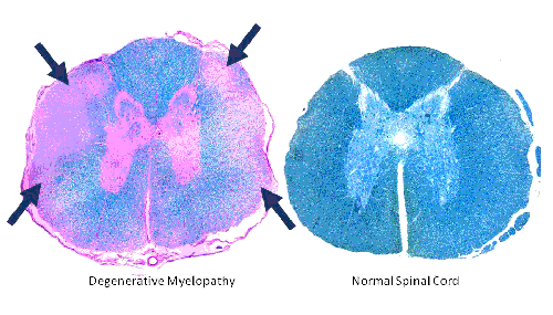
In the section of a spinal cord from a dog who has died
of DM (Left), the degeneration is seen as a loss of the
blue color at the edges (arrows) compared with the
spinal cord from a normal dog which is blue througout
(Right). |
|
This degeneration consists of both demyelination
(stripping away the insulation of these fibers) and
axonal loss (loss of the actual fibers), and interferes
with the communication between the brain and limbs.
Recent research has identified a mutation in a gene that
confers a greatly increased risk of developing the
disease.
How is degenerative myelopathy clinically diagnosed?
Degenerative myelopathy is a diagnosis of elimination.
We look for other causes of the weakness using
diagnostic tests like myelography and MRI. When we have
ruled them out, we end up with a presumptive diagnosis
of DM. The only way to confirm the diagnosis is to
examine the spinal cord under the microscope when a
necropsy (autopsy) is performed. There are degenerative
changes in the spinal cord characteristic for DM and not
typical for some other spinal cord disease.
What else can look like degenerative myelopathy?
Any disease that affects the dog’s spinal cord can cause
similar signs of loss of coordination and weakness.
Since many of these diseases can be treated effectively,
it is important to pursue the necessary tests to be sure
that the dog doesn’t have one of these diseases. The
most common cause of hind limb weakness is herniated
intervertebral disks. The disks are shock absorbers
between the vertebrae in the back. When herniated, they
can cause pressure on the spinal cord and weakness or
paralysis. Short-legged, long back dogs are prone to
slipped disks. A herniated disk can usually be detected
with X-rays of the spine and myelogram or by using more
advanced imaging such as CT scan or MRI. Other diseases
we should consider include tumors, cysts, infections,
injuries and stroke. Similar diagnostic procedures will
help to diagnose most of these diseases. If necessary,
your veterinarian can refer you to a board certified
neurologist who can aid in diagnosing degenerative
myelopathy. A directory to a neurologist near you can be
found at
American College of Veterinary
Internal Medicine
website under the "Find a Specialist Near You" link.
How do we treat degenerative myelopathy?
There are no treatments that have been clearly shown to
stop or slow progression of DM. Although there are a
number of approaches that have been tried or recommended
on the internet, no scientific evidence exists that they
work. The outlook for a dog with DM is still grave. The
discovery of a gene that identifies dogs at risk for
developing degenerative myelopathy could pave the way
for therapeutic trials to prevent the disease from
developing. Meanwhile, the quality of life of an
affected dog can be improved by measures such as good
nursing care, physical rehabilitation, pressure sore
prevention, monitoring for urinary infections, and ways
to increase mobility through use of harnesses and carts.
|
http://www.cvm.missouri.edu/neurology/dm/index.html
Guidelines for Breeding dogs who are Carrier or At Risk for
DM- DEGERERATIVE MIELOPATHY
Owners with dogs testing as Carriers (DM/N), or At-Risk
(DM/DM) are strongly encouraged to share these results with
their attending veterinarian and seek genetic counseling
when making breeding decisions.
The “DM” (mutated) allele appears to be very common in some
breeds. In these breeds, an overly aggressive breeding
program to eliminate dogs testing DM/DM or DM/N might be
devastating to the breed as a whole because it would
eliminate a large fraction of the high quality dogs that
would otherwise contribute desirable qualities to the breed.
Nonetheless, DM should be taken seriously. It is a fatal
disease with devastating consequences for the dog, and can
be a trying experience for the owners that care for them. A
realistic approach when considering which dogs to select for
breeding would be to treat the test results as one would
treat any other undesirable trait or fault. Dogs testing
At-Risk (DM/DM) should be considered to have a more serious
fault than those testing as Carriers (DM/N). Incorporating
this information into their selection criteria, breeders can
then proceed as conscientious breeders have always done:
make their breeding selections based on all the dog’s
strengths and all the dog’s faults. Using this approach and
factoring the DM test results into the breeding decisions
should reduce the prevalence of DM in the subsequent
generations while continuing to maintain and improve upon
positive, sought after traits.
We recommend that breeders take into consideration the DM
test results as they plan their breeding programs; however,
they should not over-emphasize the test results. Instead,
the test result should be one factor among many in a
balanced breeding program.
|
WHAT IS EPILEPSY?
Idiopathic epilepsy is a "diagnosis of exclusion" - there is
no test at this time that says "yes, this dog has epilepsy".
A dog experiencing repeated seizures, with no identifiable
underlying cause (tests run to exclude things that can cause
a seizure), is diagnoses as an idiopathic epileptic. Most
people don't run every test known to veterinary medicine, as
that's quite expensive and probably not productive in terms
of changing the treatment plan, but there are basic tests
that rule out major things. We have info on testing and why
to do or not do various tests in the "Basics" section of our
website - http://www.canine-epilepsy.net/. Bottom line, a
dog experiencing seizures is affected with seizures;
repeated seizures over time, the dog is called an epileptic
- but could be primary (idiopathic) or secondary epilepsy
(caused by something, such as a tumor, etc).
When we see idiopathic epilepsy in dogs in their prime -
1-5yrs - when they should be healthy and have no problems,
tests show no underlying cause, it is generally assumed they
have inherited "something" that is allowing them to seize.
That "something" is what we're trying to find. When we can
identify the mutation, or find a marker linked to the
disease, then there WILL be a test for inherited epilepsy.
We're not there yet though!
Liz Hansen
Animal Molecular Genetics Laboratory University of Missouri
- College of Veterinary Medicine
321 Connaway Hall Columbia, MO 65211
573-884-3712
HansenL@missouri.edu
|
|
The Collie Health Foundation
www.colliehealth.org
In 1986 the Collie Club Of America established the Collie
Club Of America Foundation, Inc. The concept for this
organization was conceived by the late R.L. Rickenbaugh, a
longtime breeder, along with his wife Hilda, of the
Bannerblu Collies. With the
Collie Club of
America's
assistance and cooperation, the reality of the organization
was set into motion.
The benefits would be two-fold. Not only would it provide
tax deductions for potential donors, but it would result in
additional income to the club, for some very worthwhile
causes. Previously, the Collie Club of America was only able
to give limited support for collie medical research and
related activities. Health problems, such as Collie Eye
Anomaly (CEA), Progressive Retinal Atrophy (PRA), Bloat,
Epilepsy, skin problems, Dermatomyositis (DM) and Grey
Collie Syndrome, that can affect the collie breed, need
significant funding if they are ever to be conquered. Thus,
the Foundation was born, with the primary function of
addressing the breed's major health problems. Its main
purpose is to issue grants to worthwhile organizations for
research in breeding, genetics and health issues of all dogs,
with the primary emphasis on Veterinary research as it
relates directly to the Collie. So far the Foundation has
given grants to the following areas of research: Bloat, Grey
Collie, Epilepsy, eye diseases (most notably PRA), DM and
many other health related problems. The very important
message of the Foundation is "funding research" and "education."
It is a "not-for-profit" corporation that receives its funds
through membership donations, fund raising activities and
other contributions such as $1 from every CCA member's dues.
The larger the Foundation's membership becomes, the more
generous its grants for research can be. In essence, the
Foundation has filled a large void, as prior to its
establishment, the club's commitment to medical research was
very limited. Many other breed clubs, including the
American Kennel Club,
have followed suit and established their own Health
Foundations (AKCCHF), using the CHF as the role model. The
Collie Club of America was and is a leader in this area.
Purpose & Goals of the Collie Health Foundation
Education
-
Promote appreciation and knowledge of dogs in general
and Collies in particular.
-
To further understanding of the diseases, defects,
injuries, and other ailments that afflict dogs in
general & Collies in particular.
-
To sponsor medical research on health problems,
genetics, breeding and history.
Research
-
Establish a national data base of resource materials
about Collies.
-
To sponsor medical research on health problems,
genetics, breeding and history.
Publish and Distribute Educational Materials on
-
Care
-
Treatment
-
Breeding
-
Development
-
Training
In order to meet these objectives, every year the Foundation
awards financial
Grants
to selected individuals and organizations. Periodic reports
of these research projects, are published in the
newsletter..
In its ongoing pursuit of providing the Collie fancy with
important educational and research tools, the Foundation has
sponsored book projects, such as "The Collective Writings of
Bobbee Roos." We also sponsored the two book by Kristina
Marshall, "His Dogs" and "The Lost Stories of Albert Payson
Terhune." Other health and educational materials are also
occasionally made available to the Foundation membership.
Our most recent project is a comprehensive Health Survey,
which hopefully will give us an understanding of the current
status of the breed's health. This will provide an ongoing
aid in determining our Collie health priorities when seeking
Grant funding. |
|